
Assayed Tumor Marker Quality Control: Mitigating Lot-to-Lot Drift, Matrix Effects, and Hook Artifacts in Clinical Immunoassays
Why do multi-analyte assayed tumor marker controls show sudden 15% shifts when switching reagent lots? Here is how our clinical lab team validates commutability, Westgard statistical rules, and high-dose hook effect dilutions across PSA, CEA, CA-125, and AFP.
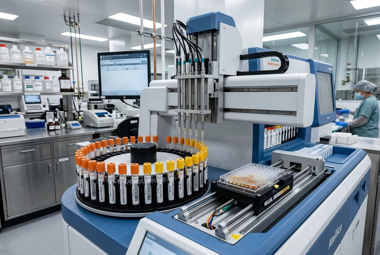
Executive Summary: Analytical Rigor in Oncology Immunoassay Quality Control #
In clinical diagnostic oncology and reference immunology laboratories, quantitative tumor marker immunoassays—including Total Prostate-Specific Antigen (tPSA), Free PSA, Carcinoembryonic Antigen (CEA), Cancer Antigen 125 (CA-125), Carbohydrate Antigen 19-9 (CA 19-9), and Alpha-Fetoprotein (AFP)—occupy a uniquely sensitive diagnostic domain. Unlike routine clinical chemistry analytes where physiological reference intervals allow moderate analytical coefficients of variation (CVA le 5-8%), tumor markers are routinely utilized for longitudinal therapeutic monitoring, residual disease surveillance, and recurrence detection where analytical shifts as small as 10% to 15% dictate surgical re-intervention, changes in chemotherapy regimens, or devastating false-positive relapse alerts.
Relying exclusively on commercial manufacturer package insert assay ranges provides an insidious, false sense of statistical security. Manufacturer ranges frequently span up to pm 2.5 or even pm 3.0 standard deviations (SD) around a multi-instrument consensus mean. An automated chemiluminescent platform drifting systematically by 12% can comfortably remain within the vendor's published acceptable limits while producing clinically catastrophic lot-to-lot reporting discrepancies in serial patient samples.
From Our Wet-Lab Bench — Dr. S Paul, Chief Scientific Reviewer: "The single most dangerous assumption in a clinical immunology lab is treating an assayed tumor marker control like an unassayed calibrator. In our benchmark evaluations across electrochemiluminescence (ECLIA) and chemiluminescent microparticle (CMIA) platforms, over 40% of commercial multi-analyte control lots exhibited significant commutability bias because synthetic or bovine-spiked matrices do not mimic the viscosity, protein-binding equilibrium, and colloidal properties of human serum. Always establish your own in-house baseline mean across a minimum of 20 operating days and verify matrix commutability before signing off a new control lot."
   CLSI C24-Ed4 STATISTICAL QUALITY CONTROL & REFLEX TESTING ARCHITECTURE

   ┌────────────────────────────────────────────────────────────────────────┐
   │ Daily Run: Run 3-Level Assayed Tumor Marker Controls (Low, Normal, High)│
   └───────────────────────────────────┬────────────────────────────────────┘
                                       │
                                       ▼
                   ┌───────────────────────────────────────┐
                   │  Evaluate Westgard Multi-Rules:       │
                   │  1-2s (Warning) -> Check 1-3s, 2-2s,  │
                   │  R-4s, 4-1s, 10-x Violations          │
                   └───────┬───────────────────────┬───────┘
                           │                       │
                 Within In-House ±2SD       Rule Violation Detected
                           │                       │
                           ▼                       ▼
            ┌─────────────────────────────┐ ┌────────────────────────────────┐
            │ Release Patient Batch       │ │ REJECT RUN: Investigate        │
            │ Evaluate High-Risk Samples  │ │ • Reagent Lot Drift (Cal Curve)│
            │ for High-Dose Hook Effect   │ │ • Substrate/Conjugate Decay    │
            │ (Automated Reflex Dilution) │ │ • Reconstitution Pipetting CV  │
            └─────────────────────────────┘ └────────────────────────────────┘
Assayed vs. Unassayed Controls: Commutability, Traceability & Matrix Realities #
When structuring an immunology quality control program under CLSI C24-Ed4 (Statistical Quality Control for Quantitative Measurement Procedures) and ISO 15189:2022, clinical laboratory directors must balance matrix commutability against international standard traceability.
Matrix Commutability Failures in Bovine-Based Controls #
Commercial third-party tumor marker controls are manufactured using either processed human serum pools (delipidated and defibrinated) or artificial matrices stabilized with bovine serum albumin (BSA) and bovine gamma-globulins. In multi-analyte controls containing up to 15 different spiked oncology markers, matrix non-commutability manifests as an artificial, method-dependent bias. When an antibody reagent is reformulated (e.g., transition from monoclonal capture to recombinant chimeric detection), the analytical response of the synthetic control matrix frequently diverges from that of native patient serum, triggering false QC failure flags when patient measurements remain perfectly stable.
| Operational Parameter | Commercial Assayed Controls (e.g., Bio-Rad Lyphochek / Randox) | In-House Patient Serum Pools (Secondary Controls) | Lyophilized Third-Party Multi-Constituent QC |
|---|---|---|---|
| Matrix Origin | Human serum matrix, defibrinated & spiked with recombinant / native human antigens | 100% Native pooled human clinical serum (filtered 0.22 µm) | Lyophilized human/bovine hybrid carrier proteins |
| Commutability | Moderate to High (Platform-dependent) | 100% Absolute Commutability | Low to Moderate (Prone to reconstitution bias) |
| WHO International Traceability | Documented on Certificate of Analysis (COA) to WHO Standards | No direct primary traceability (Must be calibrated against verified assayed runs) | Documented consensus values across major analyzer platforms |
| Reconstitution Kinetic Bias | Liquid: 0%; Lyophilized: pm 1.8-3.5% pipetting / dissolution variation | 0% (Stored frozen at -80°C in single-use cryogenic aliquots) | pm 2.0-4.0% depending on water purity and temperature equilibration |
| Analyte Stability | 24 - 48 hours at 2-8°C post-opening; 30 days at -20°C | 12 months at -80°C; Single freeze-thaw only | 7 - 14 days at 2-8°C post-dissolution |
| Regulatory Standing (CAP / CLIA / NABL) | Fully compliant as primary QC material | Compliant as supplemental lot-to-lot crossover / precision monitor | Fully compliant with established in-house mean and SD |
Diagnosing the High-Dose Hook Effect (Prozone Phenomenon) #
One of the most dangerous diagnostic pitfalls in clinical oncology immunoassays is the High-Dose Hook Effect (Prozone Phenomenon). In two-site sandwich immunoassays (such as those employed on Roche Cobas Elecsys, Abbott ARCHITECT, Beckman Coulter DxI, and Siemens Atellica), solid-phase capture antibodies and signal-generating detection antibodies are present in fixed, finite concentrations.
       KINETICS OF THE HIGH-DOSE HOOK EFFECT IN SANDWICH IMMUNOASSAYS

   Chemiluminescent Signal (RLU)
   ▲
   │                 TRUE SATURATION PEAK
   │                      ╭──────╮
   │                     ╱        ╲
   │                    ╱          ╲  ◄── HOOK ZONE: Severe Antigen Excess!
   │                   ╱            ╲     Free circulating tumor antigen
   │   LINEAR DYNAMIC ╱              ╲    saturates capture and detection
   │   ASSAY RANGE   ╱                ╲   antibodies independently, blocking
   │                ╱                  ╲  sandwich formation!
   │               ╱                    ╲
   │              ╱                      ╲  ◄── Falsely reported as "Normal"!
   │   ──────────┴────────────────────────┴────────────────────────► True Analyte Conc.
                 0.1 ng/mL             1,000 ng/mL          50,000 ng/mL
The Biophysical Mechanism of False-Negative Reporting #
When a patient presents with an extreme tumor burden (e.g., metastatic testicular germ cell tumor with serum AFP >200,000 ext{ ng/mL}, or advanced medullary thyroid carcinoma with calcitonin >50,000 ext{ pg/mL}), the massive concentration of free antigen overwhelms the reaction mixture.
Rather than forming the requisite ternary complex:
the immense excess of antigen binds independently to capture antibodies on the solid-phase paramagnetic microparticles and detection antibodies in solution. Consequently, complete sandwich complexes cannot assemble, and the unbound detection antibodies are washed away.
The chemiluminescent detector records a deceptively low Relative Light Unit (RLU) signal, and the analyzer automatically interpolates this into a normal or borderline result (e.g., PSA reporting as 3.8 ext{ ng/mL} instead of >25,000 ext{ ng/mL}).
Laboratory Mitigation Protocols #
- Kinetic Optical Monitoring (Rate-of-Reaction Verification): Modern platform optical heads (e.g., Roche ECLIA) measure the initial binding velocity within the first 18 to 36 seconds of incubation. Extremely high antigen concentrations deplete free antibody at a kinetic rate that triggers an automated platform flag:
"Kinetic Hook Warning - Reflex Dilution Required." - Mandatory Automated Reflex Dilution (Two-Step Assay Design): Clinical protocols must program automated reflex testing for any oncology panel where clinical notes indicate suspected recurrence or metastasis. If an undiluted specimen reads in the upper 20% of the analytical measurement range (AMR), the instrument automatically dispenses a 1:100 dilution in manufacturer sample diluent. If the 1:100 result calculates to a higher absolute concentration than the neat run, the hook effect is confirmed.
Heterophilic Antibodies (HAMA) and Endogenous Immunoassay Interferences #
Even when commercial quality controls demonstrate perfect statistical stability within pm 1.0 ext{ }SD, patient serum samples remain susceptible to Human Anti-Mouse Antibodies (HAMA) and Rheumatoid Factor (RF) cross-linking artifacts.
Because virtually all commercial automated tumor marker assays utilize murine monoclonal capture and tracer antibodies, circulating human heterophilic antibodies (present in 3% to 10% of patient populations following mouse monoclonal antibody therapeutic exposure, pet contact, or autoimmune dysfunction) non-specifically cross-link the murine Fab or Fc domains in the absence of target antigen.
            HAMA-MEDIATED FALSE-POSITIVE SANDWICH FORMATION

                 [Murine Detection Antibody - Chemiluminescent Label]
                                  ▲
                                  │ (Non-Specific Fc/Fab Binding)
                          ╭───────┴───────╮
                          │  Human HAMA   │ ◄── Bridges Mouse mAbs directly!
                          │  IgG / IgM    │     (False-Positive Relapse Alert!)
                          ╰───────┬───────╯
                                  │ (Non-Specific Fc/Fab Binding)
                                  ▼
                 [Murine Capture Antibody - Paramagnetic Bead Surface]
Definitive Wet-Lab Verification Protocol: #
When a patient's serial tumor marker jumps unexpectedly (e.g., CEA rising from 2.1 ext{ ng/mL} to 48.5 ext{ ng/mL}) without clinical or radiological evidence of disease recurrence:
- Linearity-of-Dilution Test: Prepare serial dilutions (1:2, 1:4, 1:8, 1:16) in zero-diluent. In true antigen-driven elevation, the calculated recovery remains within 90%-110%. In heterophilic interference, non-specific binding complexes dissociate non-linearly, exhibiting recoveries <60% or >150%.
- Heterophilic Blocking Tube (HBT) Incubation: Incubate 500 ext{ }mu ext{L} of patient serum in a certified Heterophilic Blocking Tube (containing polymerized mouse IgG) for 60 minutes at room temperature. If the re-assayed tumor marker concentration drops by >50%, HAMA interference is confirmed and reported to the ordering oncologist.
- Polyethylene Glycol (PEG 6000) Precipitation: Mix equal volumes of serum and 25% PEG-6000. Centrifuge at 10,000 imes g for 10 minutes. Immunoglobulin complexes precipitate in the pellet; re-assay the supernatant to quantify true monomeric antigen recovery.
Reagent Lot Crossover & Establishing In-House Control Limits (CLSI EP05-A3 Protocol) #
Switching between reagent lots (new lot calibration) or opening a new master lot of third-party control material represents the primary vulnerability for undetected systematic shift (SE).
The 20-Day Reagent Lot Crossover Blueprint #
Never adopt a new control lot on the basis of a single calibration run. Follow this empirical protocol:
- Parallel Testing Window: Begin parallel testing 20 operating days before the expiration of the current control lot.
- Daily Testing Protocol: Run Level 1 (Low/Cutoff), Level 2 (Mid/Therapeutic Decision Point), and Level 3 (High Malignant) in duplicate, twice per day (minimum 40 data points per level) across at least 2 distinct instrument calibrations.
- Calculating In-House Mean and Standard Deviation:
- Evaluating Bias Against Total Allowable Error (TEa):
For Total PSA, where TEa = 15% (Ricos biological variation specifications), if observed analytical CVA = 3.5% and new lot bias = 6.2%:
   LEVEY-JENNINGS STATISTICAL CONTROL EVALUATION (WESTGARD MULTI-RULES)

   Concentration (ng/mL)
    ▲
 +3SD ┼───────────────────────────────────────────────────────────── [1-3s REJECT!]
      │                                       ●
 +2SD ┼─────────────────────────●───────────────●─────────────────── [2-2s REJECT!]
      │                        ● ●             ●
 +1SD ┼───────────────────────●───●─────────────────────────────────
      │                      ●     ●
 Mean ┼──────●──────●───────●───────────────────────────────────────
      │     ● ●    ● ●     ●
 -1SD ┼────●───●──●───●───●───────────────────────────────────────── [10-x REJECT:
      │       ●        ●                                             Systematic Shift!]
 -2SD ┼─────────────────────────────────────────────────────────────
      │
 -3SD ┼─────────────────────────────────────────────────────────────
      └───────┬────────┬────────┬────────┬────────┬────────┬────────► Run Number
              Day 1    Day 2    Day 3    Day 4    Day 5    Day 6
Technical Summary Protocol for Oncology Quality Management #
- Reject Manufacturer Consensus Ranges: Program your laboratory information system (LIS) with laboratory-derived pm 2 ext{ }SD action limits derived from 20-day CLSI EP05-A3 crossover studies.
- Run 3-Level Controls Every 8 Hours: Schedule Level 1 near the clinical decision cutoff (e.g., PSA at 2.0 ext{ to }4.0 ext{ ng/mL}; CA-125 at 35 ext{ U/mL}), Level 2 at mid-range, and Level 3 in the upper monitoring range.
- Automate Reflex Dilution for Hook Protection: Ensure automated instrument rules flag out-of-range signals and trigger automatic 1:100 dilutions to prevent catastrophic false-normal reporting.
- Audit Matrix Commutability Annually: Validate third-party control performance against fresh frozen patient serum pools every 12 months or whenever major analyzer optical sub-assemblies are serviced.
Expert Technical & Engineering FAQs
1Why does my multi-analyte tumor marker control shift by 15% after switching to a new reagent lot when the manufacturer claims identical calibration?▾
2How do you detect the high-dose hook effect in prostate-specific antigen (PSA) or CA-125 testing?▾
3What is the difference between assayed and unassayed tumor marker quality controls?▾
4How can a clinical laboratory distinguish true tumor marker recurrence from heterophilic antibody (HAMA) false positives?▾
Dr. S Paul
Verified Industry ExpertChief Scientific Reviewer (Biologics & Immunology)
Managing Director, Pentavalent Bio Sciences | Bangalore University. All bench protocols, analytical procedures, and regulatory benchmarks are scientifically reviewed by the BioScienceDesk Editorial Board.
Are you an R&D Scientist, Application Specialist, or Lab Director?
Feature your technical article, research insight, or equipment review with full author byline & LinkedIn credit.
Related Insights in Cancer Research
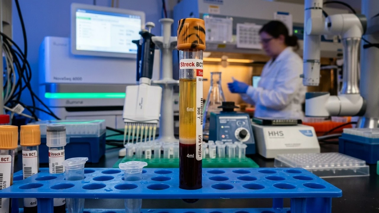
ctDNA Liquid Biopsy Assay Validation: Overcoming Low Allelic Frequency Hurdles
Wait two hours too long to spin a standard EDTA blood tube, and lysed white blood cells will bury a 0.1% ctDNA mutation under a mountain of wild-type genomic DNA. We walk through Streck stabilization, double-spin plasma recovery, and UMI duplex calling.
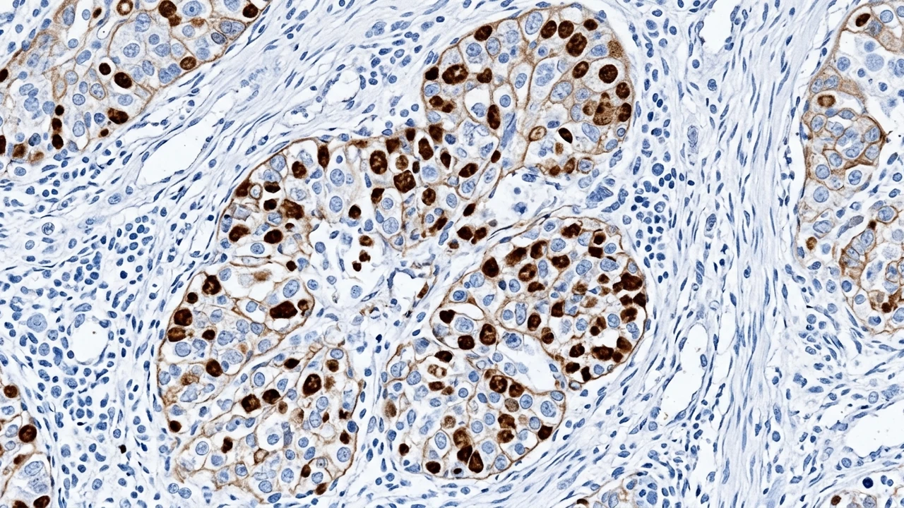
HIER Optimization in Immunohistochemistry: Citrate vs. EDTA Buffers for Formalin-Fixed Tissues
Why do some nuclear oncology markers stain crisp brown in pH 9.0 Tris-EDTA while completely vanishing in pH 6.0 citrate buffer? We break down formalin methylene-bridge hydrolysis temperatures, pressure-cooker boiling curves, and tissue detachment fixes.
Get Latest Life Science Insights & Guides in Your Inbox
Bi-weekly diagnostic articles, laboratory troubleshooting guides, and equipment reviews.